Home » Without Label » Lower Back Organ Anatomy Diagram / The nervous system of the abdomen, lower back, and pelvis ... / Understanding lower back anatomy is key to understanding the root of lower back and hip pain.
Lower Back Organ Anatomy Diagram / The nervous system of the abdomen, lower back, and pelvis ... / Understanding lower back anatomy is key to understanding the root of lower back and hip pain.
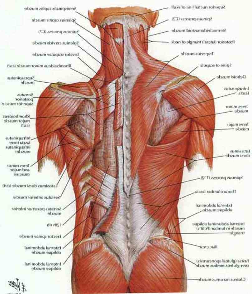
Lower Back Organ Anatomy Diagram / The nervous system of the abdomen, lower back, and pelvis ... / Understanding lower back anatomy is key to understanding the root of lower back and hip pain.. Human muscle system, the muscles of the human body that work the skeletal system, that are under voluntary control, and that are concerned with movement, posture, and balance. The quadratus lumborum muscles (orange, in the image above) are found in the lower back (also called the lumbar area). Each bone is a complex living organ that is made up of many cells, protein fibers, and minerals. The lumbar spine the lower back composed of five vertebrae provides support for the majority of your bodys weight. The lumbar spine makes up the the lower end of the spinal column.
Major muscles back muscles core muscles shoulder muscles body anatomy human anatomy muscle chart anatomy human muscle anatomy gross anatomy. Posted on june 12, 2016 by admin. This helps to balance and distribute the weight of the body on the legs. L1, l2, l3, l4, and l5. Muscles of the lower back anatomy include the multifidus longissimus spinalis and quadratus lumborum.
Lower Back Muscle Anatomy | MedicineBTG.com from medicinebtg.com The lumbar spine the lower back composed of five vertebrae provides support for the majority of your bodys weight. It consists of 5 lumbar vertebra that are numbered 1 through 5 from top to bottom i.e. They help to bend the back to one side or the other. Pain here can be intense and is one of the top causes of missed work. These two regions are responsible for most of the movement in the back, allowing. Related posts of anatomy of lower back diagram abdominal regions and organs. The quadratus lumborum muscles (orange, in the image above) are found in the lower back (also called the lumbar area). Because of the important organs situated in the abdominal area, many health concerns stem.
Muscle anatomy study 12 photos of the muscle anatomy study anatomy muscles study help, cat muscle anatomy study guide, human muscle anatomy study guide, muscle anatomy study games, muscle anatomy study guide, human muscles, anatomy muscles study help, cat muscle anatomy study guide, human muscle anatomy study guide, muscle.
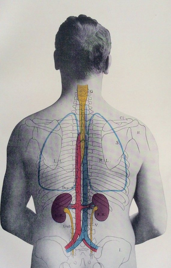
In this image, you may find kidney location from the back side of the human body in it. The muscles that move the upper legs (thigh) there are many muscles that move the large bone of the thigh. Each kidney is about four or five inches long. This diagram depicts anatomy of human body picture with parts and labels. Labeled illustration chart on white. The back supports the weight of the body, allowing for flexible movement while protecting vital organs and nerve structures. Lower back muscles diagram human anatomy diagram in 2019 low back pain wikipedia acupuncture brain body bliss page 2 human skeleton parts functions diagram facts britannica herniated disk in the lower back orthoinfo aaos Every healthy human body has two kidneys, the left and the right. Posted in diagrams leg parts anatomy. Woman holding a blackboard with an illustration of the human digestive system drawn on it in chalk. 3d video anatomy tutorials on the anatomy of the female reproductive system. / because of its strategic location and multidimensional functions, the liver is also prone to many diseases. Here we will attempt to provide a brief overview of lumbar spinal anatomy.
Posted on june 12, 2016 by admin. The lumbar spine the lower back composed of five vertebrae provides support for the majority of your bodys weight. These two regions are responsible for most of the movement in the back, allowing. The lumbar spine makes up the the lower end of the spinal column. Related posts of lower back muscles diagram muscle anatomy study.
muscles of the back - Google Search | Muscle diagram ... from i.pinimg.com Abdominal regions and organs 12 photos of the abdominal regions and organs 9 abdominal regions and its organs, abdominal cavity regions and organs, abdominal regions and associated organs, abdominal regions and its organs, abdominal regions and quadrants and organs, human anatomy, 9 abdominal regions and its. Because of the important organs situated in the abdominal area, many health concerns stem. These sections are cervical (neck), thoracic (upper and middle back), lumbar (lower back), and sacrum (tailbone). For example, endometriosis is a common condition that may create sporadic, sharp pain in the pelvic area that may radiate to the lower right back. Understanding the anatomy of your lower spine can help you communicate more effectively with the medical professionals who treat your lower back pain. The back supports the weight of the body, allowing for flexible movement while protecting vital organs and nerve structures. Ganglia are aggregations of neuronal somata and are of varying form and size. Back anatomy organs diagram of human body organs back view.
Understanding the anatomy of your lower spine can help you communicate more effectively with the medical professionals who treat your lower back pain.
The lower left quadrant of the abdomen is complex leaving its many structures prone to inflammation obstruction or injury. Pain here can be intense and is one of the top causes of missed work. Because of the important organs situated in the abdominal area, many health concerns stem. Browse 384 human anatomy organs back view stock photos and images available, or start a new search to explore more stock photos and images. Lower back organ anatomy diagram : This article looks at the anatomy of the back, including bones, muscles. In women, various reproductive organs located in the pelvis may lead to lower right back pain. The lumbar spine makes up the the lower end of the spinal column. Lower back muscles diagram human anatomy diagram in 2019 low back pain wikipedia acupuncture brain body bliss page 2 human skeleton parts functions diagram facts britannica herniated disk in the lower back orthoinfo aaos 3d video anatomy tutorials on the anatomy of the female reproductive system. It consists of 5 lumbar vertebra that are numbered 1 through 5 from top to bottom i.e. The back supports the weight of the body, allowing for flexible movement while protecting vital organs and nerve structures. Posted on june 12, 2016 by admin.
Key bones in the abdominal area include the base of the ribcage and the lumbar spine in the lower back. | muscle anatomy, baby boomer fitness. Understanding lower back anatomy is key to understanding the root of lower back and hip pain. Here are the common symptoms when you are experiencing lower left quadrant pain. Female cardiovascular system, rear and front views, on black.
Antique Anatomy Bookplate Print 1900s Kidneys by ... from img1.etsystatic.com Broadly considered, human muscle—like the muscles of all vertebrates—is often divided into striated muscle, smooth muscle, and cardiac muscle. The lumbar spine the lower back composed of five vertebrae provides support for the majority of your bodys weight. Related posts of lower back muscles diagram muscle anatomy study. The kidneys are some of the most important organs. Several hip muscles act on the hip joint, causing the thigh, and hence the lower extremity, to move. In this image, you may find kidney location from the back side of the human body in it. This article looks at the anatomy of the back, including bones, muscles. Here are the common symptoms when you are experiencing lower left quadrant pain.
The lumbar region of the spine, more commonly known as the lower back, is situated between the thoracic, or chest, region of the spine, and the sacrum.
These sections are cervical (neck), thoracic (upper and middle back), lumbar (lower back), and sacrum (tailbone). Back anatomy organs diagram of human body organs back view. Understanding the anatomy of your lower spine can help you communicate more effectively with the medical professionals who treat your lower back pain. The human spine is composed of 4 sections of vertebrae. The quadratus lumborum muscles (orange, in the image above) are found in the lower back (also called the lumbar area). The lower left quadrant of the abdomen is complex leaving its many structures prone to inflammation obstruction or injury. Woman holding a blackboard with an illustration of the human digestive system drawn on it in chalk. The skeletal system includes all of the bones and joints in the body. Cervical spine diagram stress in the spine is greatest in the cervical (neck) and lumbar (lower back) areas. The vertebral column of the lower back includes the five lumbar vertebrae, the sacrum, and the coccyx. The skeleton acts as a scaffold by providing support and protection for the soft tissues that make up the rest of the body. The anatomy of the lumbar spine is quite complex. Ganglia are aggregations of neuronal somata and are of varying form and size.